
Resuscitation
Learner monitor, instructor console, cases, your own scenarios and a report for the debriefing.
REASEL · educational software
Vital signs monitor, electrocardiogram, blood gas analysis and ultrasound for teaching resuscitation, on Windows or Mac. One-time payment in US dollars; you receive your code and the download link by email.
For educational use; it is not a medical device. Pulso REASEL simulates signals for teaching and must not be used to monitor or diagnose patients.
Beta version. Special price for early users. Tell us about your experience at contacto@reaselatinoamerica.com: it helps us improve it.
The suite
We build every part of Pulso REASEL at REASEL. The monitor, the ECG, the blood gas and the ultrasound read the same patient: what changes in the case changes in all of them.
Learner monitor, instructor console, cases, your own scenarios and a report for the debriefing.
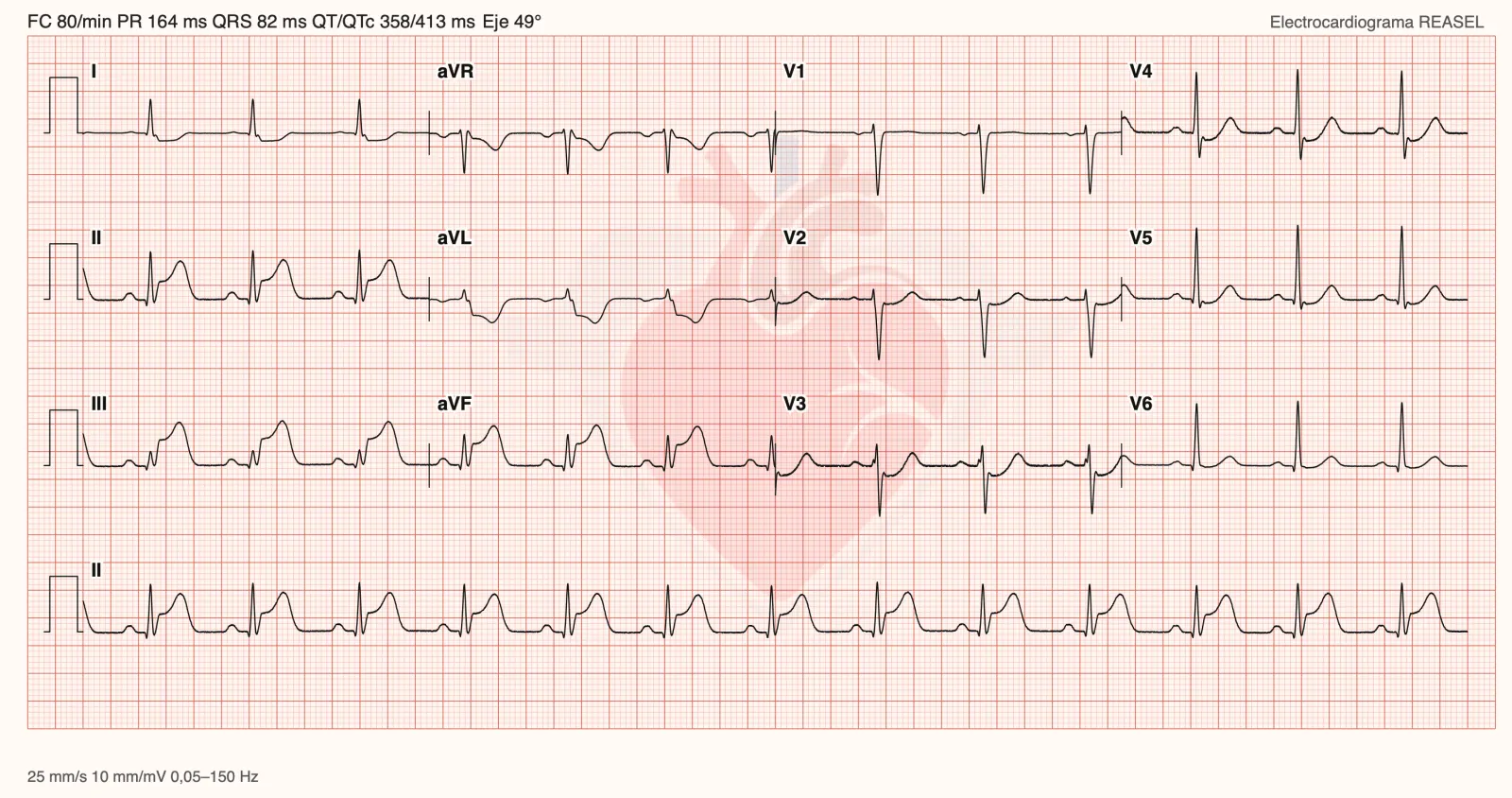
Twelve leads on graph paper, generated by Electrocardiograma REASEL.
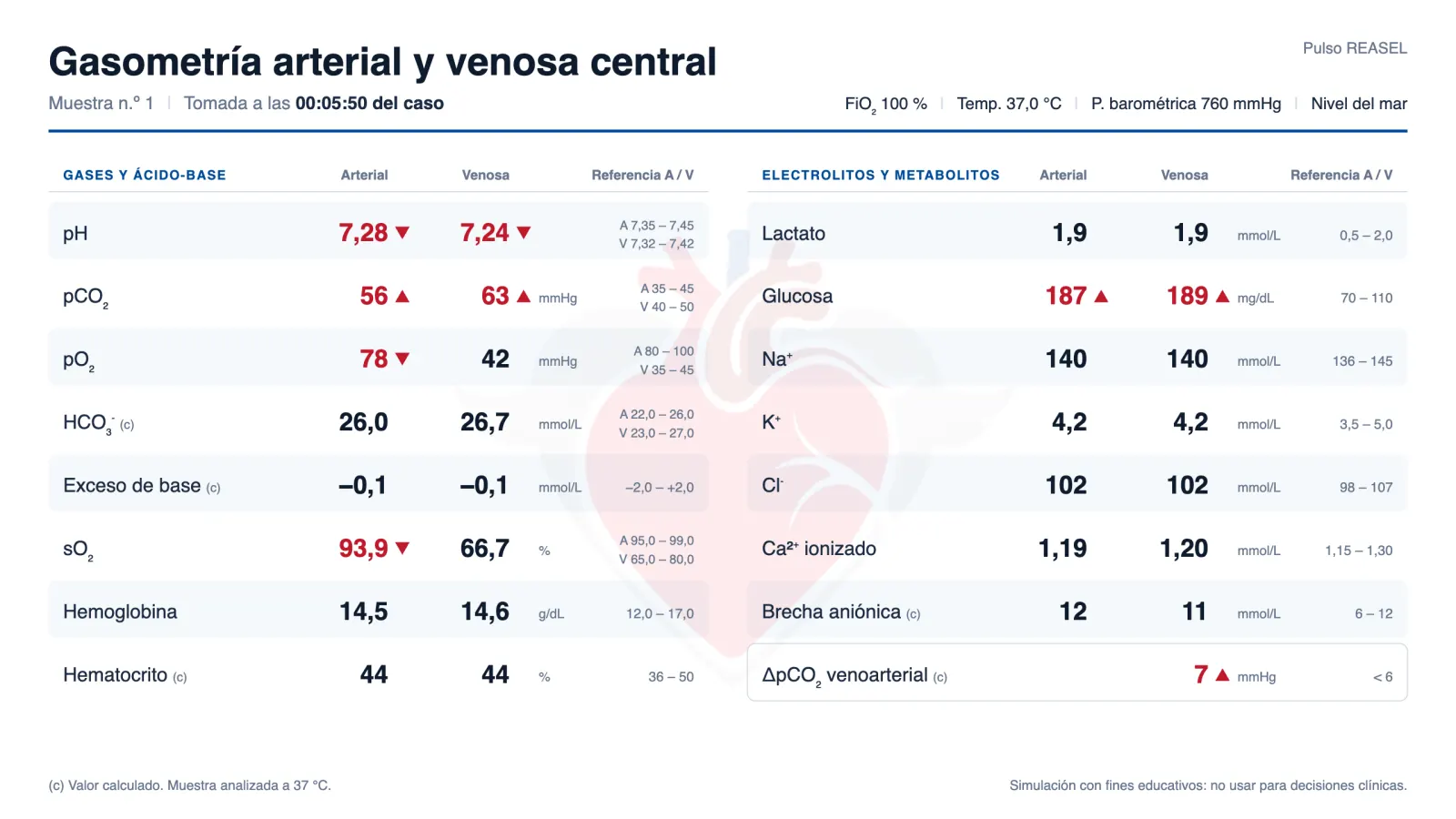
Arterial and venous blood gas and central lab results that match the patient’s condition at that moment.
Lung, heart, vena cava and venous congestion with color Doppler, moving in time with the case.
Pressure-volume loops of both ventricles, 13 patients and 11 drugs. Connected to the monitor, the blood gas and the ultrasound.
Chest X-ray. Not available yet.
What it does
Start the case, change the rhythm and vital signs, and record what the team does. The same screen shows what the learner is seeing.
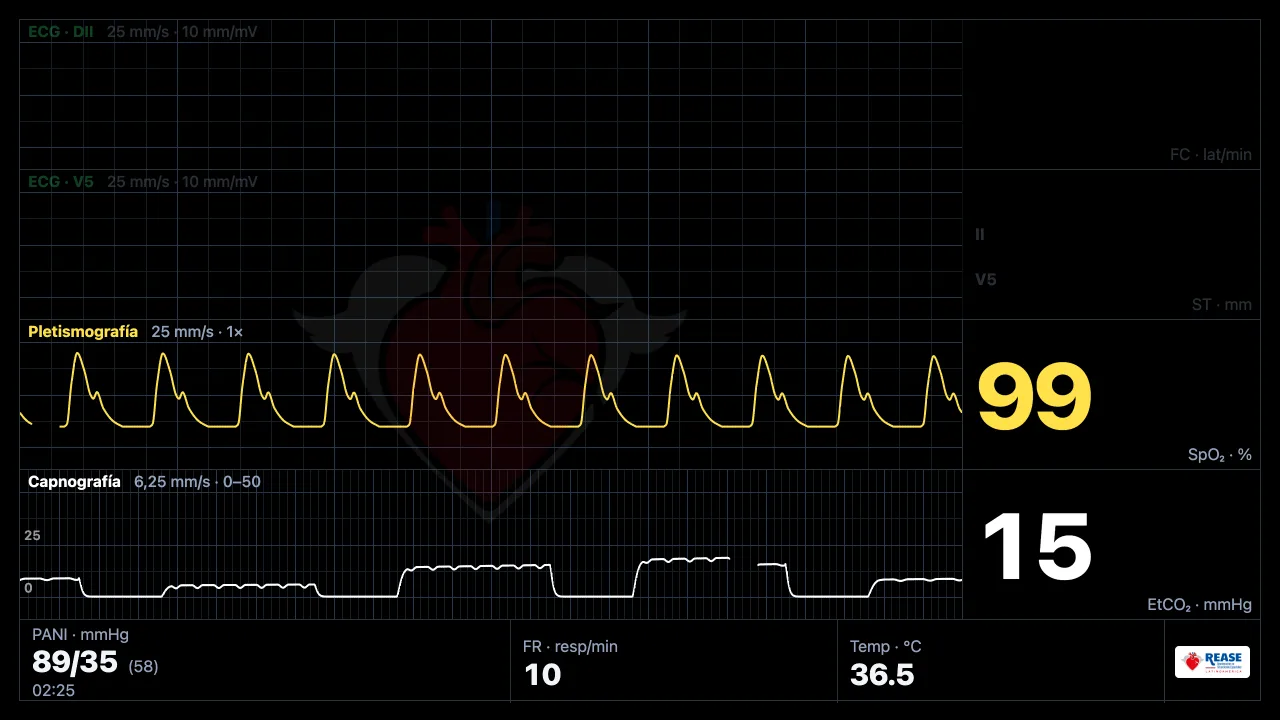
The learner sees a full-screen monitor with ECG, plethysmography, capnography and blood pressure. The rhythm changes when the case calls for it.
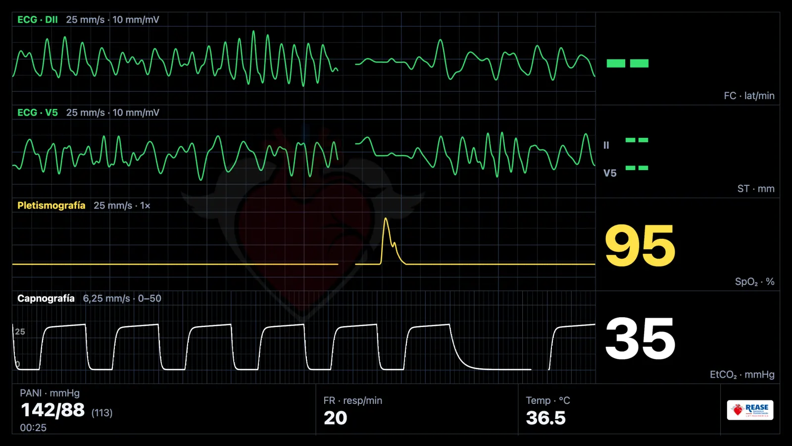
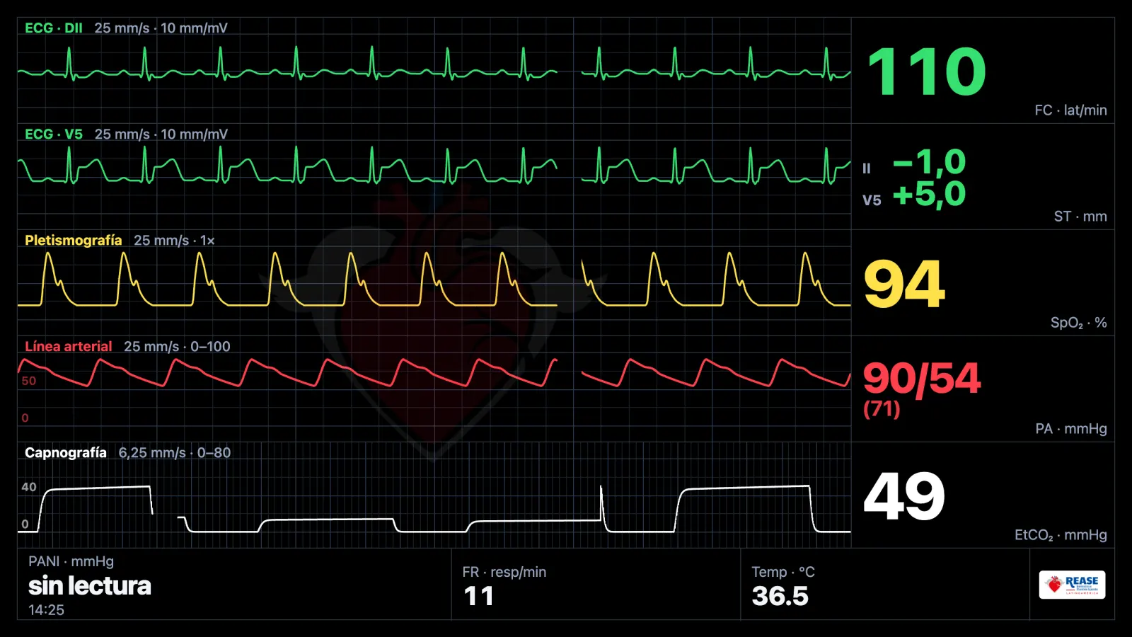
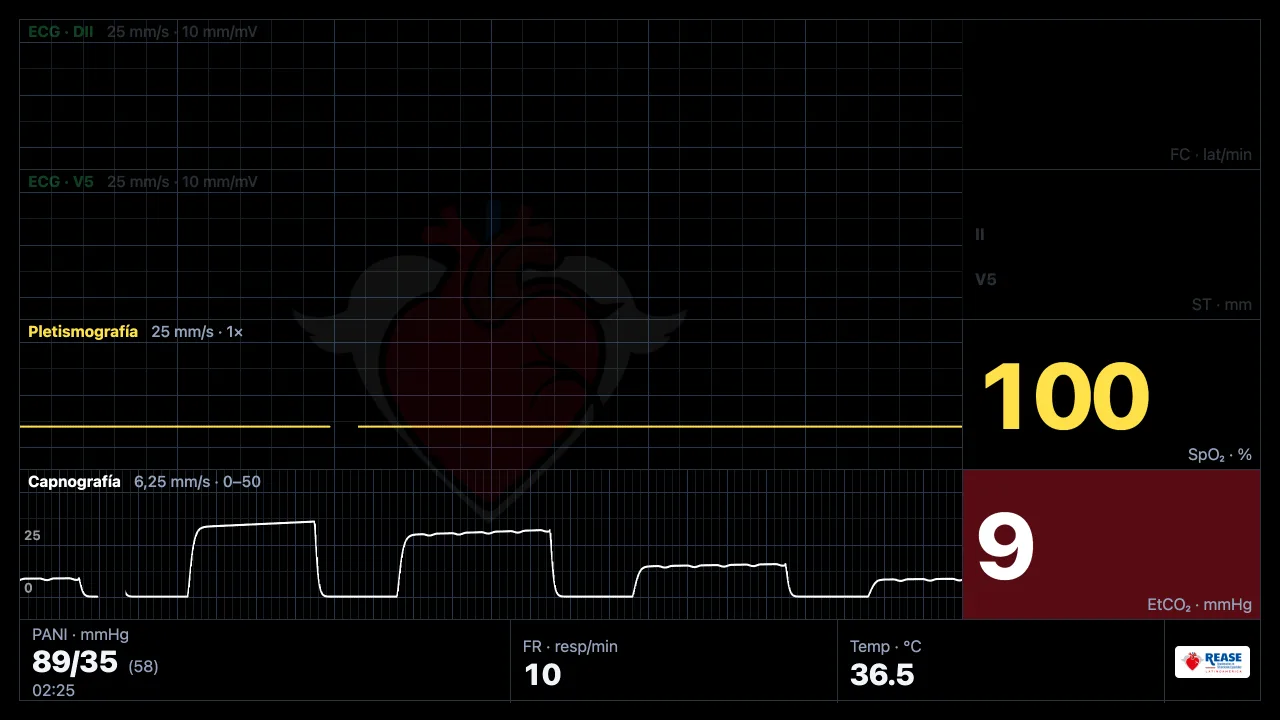
The instructor marks CPR as good or poor, or adjusts the rate, depth and chest recoil. With poor CPR the end-tidal CO₂ drops and the pulse wave is lost; with good CPR they come back.
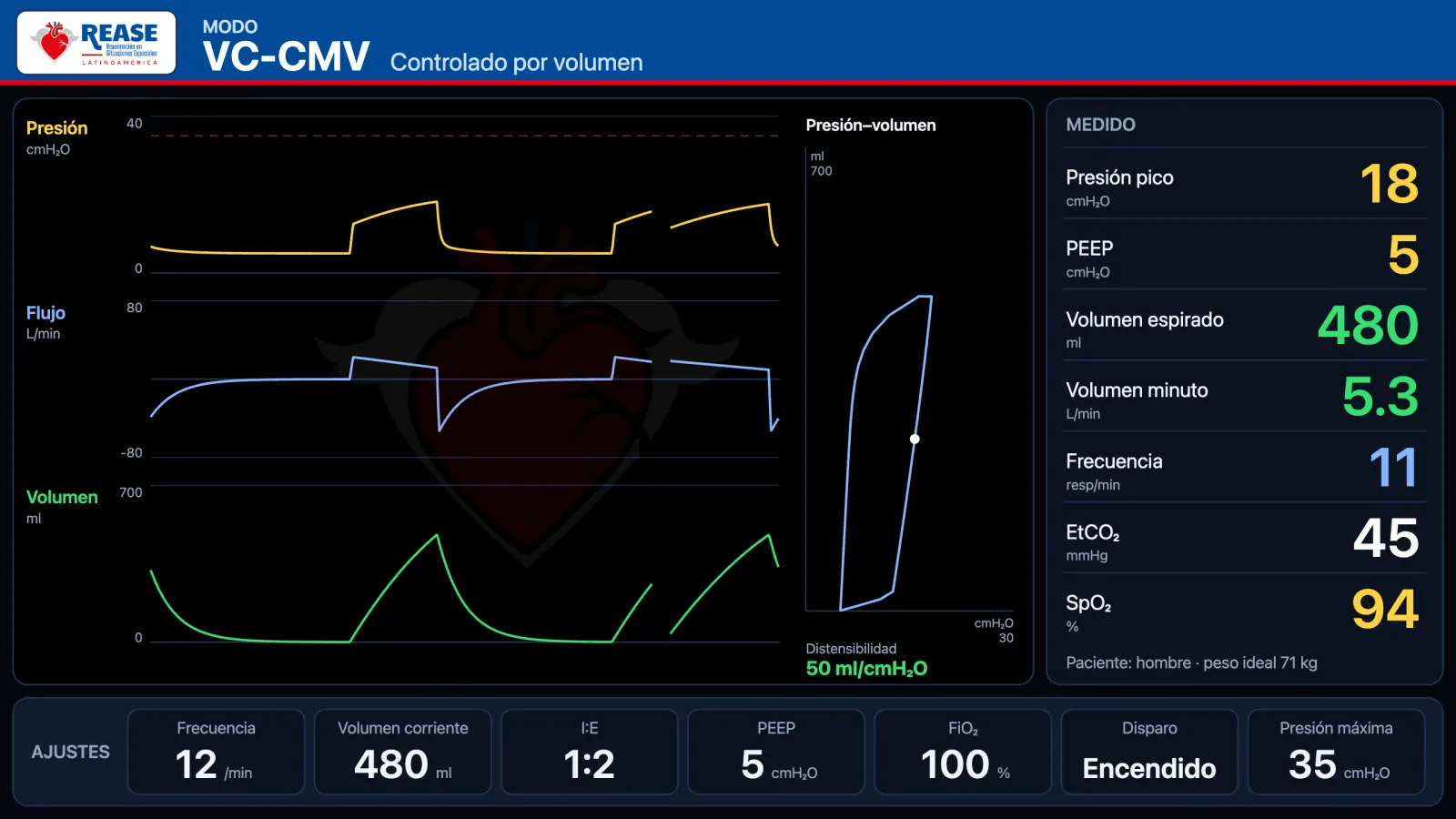
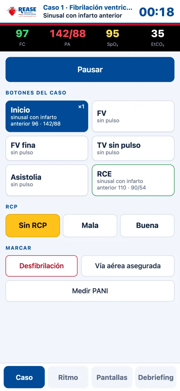
A second screen shows the ventilator, the ECG or the blood gas. The instructor can also run the case from a phone: it pairs with a QR code, with no app to install.
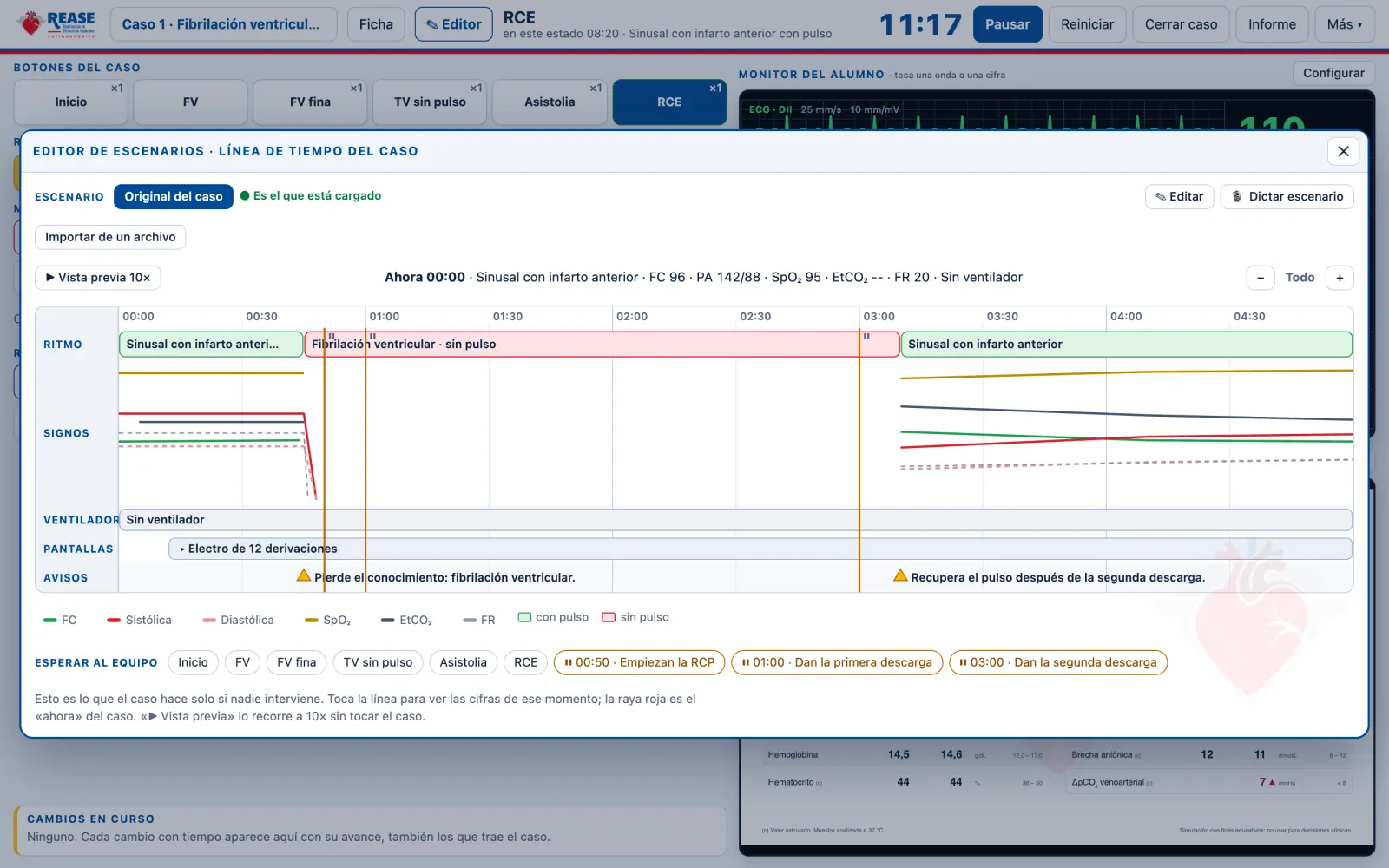
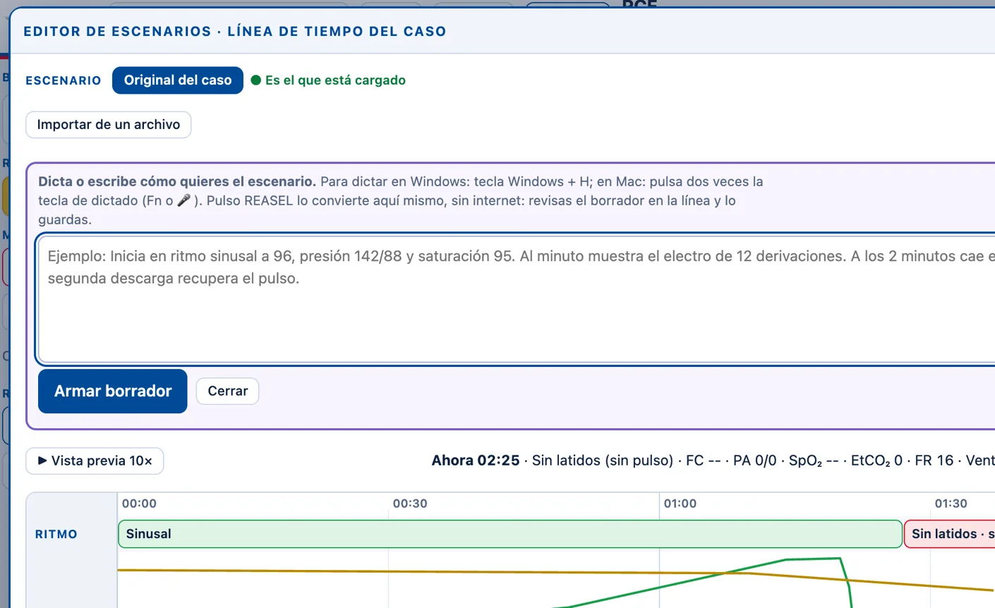
Build your cases as you would in a video editor: drag blocks for rhythm, vital signs, ventilator, screens and prompts onto the timeline. Or dictate the scenario in your own words and Pulso REASEL drafts it right there, with no internet.
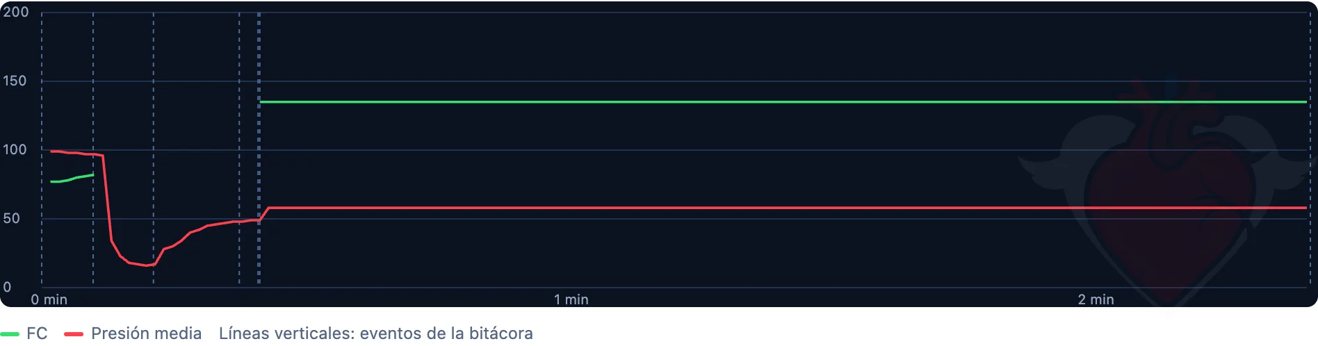
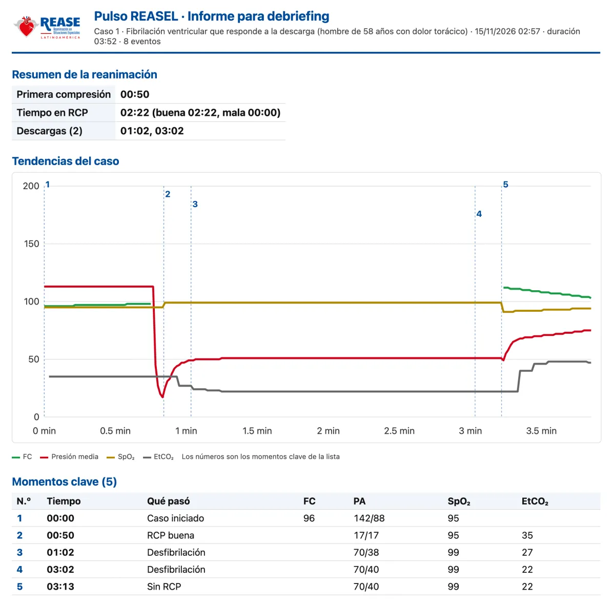
During the case you see the heart rate and blood pressure trends, with each event marked. At the end, the report brings together the resuscitation summary, the trends and the key moments.
Tests and imaging
Generated by Electrocardiograma REASEL, our own engine, on ECG grid paper.
The results sheet appears on the second screen and matches the patient’s condition at that moment.
The image moves live, in time with the heart and respiratory rate of the case, and the instructor chooses the finding.
For venous congestion, the image carries color Doppler and the pulsed Doppler tracings of the hepatic vein, the portal vein and the renal veins. There is also the liver with the right kidney, with free fluid.
Included, with continuous updates
The why behind the numbers on the monitor. A patient with a complete circulation that responds beat by beat: you change the volume, resistance, contractility or heart rate, give a drug, and the class sees what happens and why.
Live, alongside cardiac function and venous return on a single graph.
Arterial and mean pressure, heart rate, cardiac output, left and right ejection fraction, central venous pressure, pulmonary artery, wedge pressure and stroke volume.
Healthy, frail, septic, heart failure, hypovolemia, aortic stenosis, mitral regurgitation, right heart failure, pulmonary embolism, tamponade, tension pneumothorax and more.
Propofol, phenylephrine, ephedrine, esmolol and atropine as boluses; norepinephrine, epinephrine, vasopressin, dobutamine, milrinone and nitroglycerin as infusions.
Pulmonary resistance, pericardial effusion, pressure inside the chest and obstruction of venous return.
The instructor can blunt it, as happens under anesthesia.
The same patient drives the case monitor, passes its cardiac output to the blood gas and its measurements to the cardiac ultrasound on the second screen.
It is our own model for teaching physiology; the figures are illustrative and do not guide dosing. For now, in the Spanish edition.
Licenses
Pulso REASEL for Windows or Mac.
open the program, choose “Activate” and enter your code (an internet connection is needed that one time).
after that it works offline.
Clinical validation
The electrocardiograms in Pulso REASEL are generated by Electrocardiograma REASEL, our own engine. To confirm that they are recognizable and useful for teaching, cardiologists review them blind, mixed with real reference ECGs.
The review is under way. The cardiologists who take part and authorize their name will be listed here.
Being listed as a reviewer is an acknowledgment and implies no responsibility for the program’s content or its use.
We are working on the ultrasound with the Wiggers diagram and on the chest X-ray. They are not part of the program yet.